
 Purpose / What It Accomplishes #
Western blotting, also known as immunoblotting, is a widely used and powerful analytical technique in molecular biology and proteomics. Its primary purpose is to detect and semi-quantify a specific protein of interest from a complex mixture of proteins extracted from cells or tissues. It also provides information about the protein’s apparent molecular weight and can identify post-translational modifications.63
Principle / Theoretical Basis #
Western blotting is a multi-step process that combines gel electrophoresis with antibody-based detection. The principle relies on separating proteins by size, transferring them to a solid support, and then using specific antibodies to identify the target protein.
- Gel Electrophoresis (SDS-PAGE): Proteins are first denatured (unfolded) and coated with the anionic detergent sodium dodecyl sulfate (SDS), which imparts a uniform negative charge proportional to their length. This ensures that when separated by polyacrylamide gel electrophoresis (SDS-PAGE), proteins migrate through the gel primarily based on their molecular weight.64
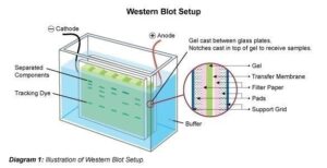
- Protein Transfer (Blotting): After separation, the proteins are electrically transferred from the gel onto a solid membrane support (typically nitrocellulose or polyvinylidene difluoride (PVDF)). The membrane binds the proteins, immobilizing them in their separated positions.64
- Blocking: The membrane’s remaining non-specific binding sites are blocked with a protein solution (e.g., nonfat milk or bovine serum albumin) to prevent non-specific binding of antibodies in subsequent steps.64
- Antibody Incubation: The membrane is then incubated with a primary antibody that is highly specific for the target protein. After washing, a secondary antibody, conjugated to an enzyme (e.g., horseradish peroxidase (HRP) or alkaline phosphatase (AP)) or a fluorescent dye, is added. This secondary antibody recognizes and binds to the primary antibody.64
- Detection: The enzyme conjugated to the secondary antibody reacts with a specific substrate to produce a detectable signal (e.g., chemiluminescence, colorimetric precipitate, or fluorescence). The intensity of this signal is proportional to the amount of target protein present.64
Step-by-Step Explanation #
- Equipment and Reagents Required: Gel electrophoresis apparatus (for SDS-PAGE), power supply, protein-binding membranes (nitrocellulose or PVDF), filter paper and fiber pads for creating the gel-membrane sandwich, a protein transfer (blotting) apparatus, rocking platform/shaker, detection system (e.g., film, charged-coupled device (CCD) camera for chemiluminescence, or fluorescent imager), various buffers (lysis buffer, SDS-PAGE running buffer, transfer buffer, wash buffers like TBS-T or PBS-T), blocking reagents (nonfat dry milk, BSA), primary antibody, secondary antibody (enzyme- or fluorophore-conjugated), and detection reagents (e.g., enhanced chemiluminescence (ECL) substrate, fluorescent dyes, chromogenic substrates).63
- Workflow from Start to Finish:
- Sample Preparation: Cells or tissues are lysed using appropriate lysis buffers (e.g., RIPA, NP-40) containing protease and phosphatase inhibitors to extract proteins and prevent degradation. Samples are then denatured by heating in a sample buffer containing SDS and a reducing agent (e.g., DTT or β-mercaptoethanol).64
- Gel Electrophoresis (SDS-PAGE): The denatured protein samples are loaded into wells of a polyacrylamide gel. A protein ladder (marker of known molecular weights) is also loaded. An electric current is applied, causing proteins to migrate through the gel and separate by size.64
- Protein Transfer (Blotting): After electrophoresis, the gel is placed in contact with a protein-binding membrane and a stack of filter papers/pads. This “sandwich” is then placed in a transfer apparatus, and an electric current is applied to drive the proteins from the gel onto the membrane, where they become immobilized.64 Transfer efficiency can be confirmed using stains like Ponceau S.64
- Blocking: The membrane is incubated in a blocking solution (e.g., 5% nonfat dry milk or BSA in Tris-buffered saline with Tween-20 (TBST) or phosphate-buffered saline with Tween-20 (PBST)) to cover all unoccupied binding sites, preventing non-specific antibody binding.64
- Primary Antibody Incubation: The membrane is incubated with the primary antibody, diluted in blocking solution, which specifically recognizes and binds to the target protein. Incubation can range from 1 hour at room temperature to overnight at 4°C.64
- Washing: The membrane is washed multiple times with wash buffer (e.g., TBST) to remove unbound primary antibody.
- Secondary Antibody Incubation: The membrane is then incubated with a secondary antibody, conjugated to an enzyme or fluorophore, which recognizes the primary antibody (e.g., anti-mouse IgG for a mouse primary antibody). This step provides signal amplification.64
- Washing: The membrane is washed multiple times to remove unbound secondary antibody.
- Detection:
- Chemiluminescent Detection: For HRP-conjugated secondary antibodies, a chemiluminescent substrate (e.g., ECL reagents containing luminol and peroxide) is added. The enzyme catalyzes a reaction that produces light, which is then detected by exposure to X-ray film or a CCD camera.64
- Fluorescent Detection: For fluorophore-conjugated secondary antibodies, the membrane is directly imaged using a fluorescent imager that excites the fluorophore and detects its emission.64
- Chromogenic Detection: For AP or HRP, a chromogenic substrate produces a colored precipitate directly on the membrane.64
- Analysis: The detected bands are analyzed for molecular weight (by comparison to the ladder) and signal intensity (for semi-quantification). Loading controls (housekeeping proteins like beta-actin or alpha-tubulin) are often used to normalize protein loading across lanes.63
Variations / Modifications #
- Detection Systems: Western blotting can employ chromogenic detection (producing a colored precipitate, long-lasting signal but difficult for multiplexing), fluorescent detection (emitting light, easier for multiplexing and co-localization but susceptible to photobleaching), or chemiluminescent detection (producing light, highly sensitive).64
- Signal Amplification Methods: Various methods enhance signal, including the Avidin-Biotin Complex (ABC) method, Labeled Streptavidin Biotin (LSAB) method, and Polymer-based methods (e.g., EnVision, ImmPRESS systems), which attach multiple enzyme molecules to secondary antibodies for increased sensitivity and reduced background.65
- Transfer Methods: Wet transfer (submerged in buffer) is common, while semi-dry transfer uses less buffer.
- Total Protein Normalization: Newer methods like Ponceau S staining or Stain-Free gels are increasingly used for normalization instead of housekeeping proteins, which can vary under experimental conditions.64
Applications #
Western blotting is widely applied in scientific and clinical procedures. Its most common uses include detecting the presence of a specific protein in complex biological mixtures, semi-quantifying relative protein levels between different samples, and evaluating protein expression levels in cells.64 It is invaluable for identifying post-translational modifications (PTMs) such as phosphorylation, ubiquitination, and glycosylation, which are crucial for understanding protein function and cellular signaling pathways.64 Western blotting also plays a role in confirming protein folding, conformational changes, and stability, and is often used as a verification step after protein purification or in diagnostic settings for specific protein biomarkers.
Strengths and Limitations #
- Strengths: Western blotting is a powerful and widely adopted technique capable of specific detection and semi-quantification of individual proteins. It provides information about the protein’s molecular mass, which is an advantage over other antibody-based methods like ELISA. Modern detection methods offer high sensitivity, and fluorescent detection enables multiplexing (detecting multiple targets simultaneously).64 It is generally more cost-effective than mass spectrometry for targeted protein detection.64
- Limitations: Western blotting is a delicate and time-consuming process, typically taking 2-3 days, with many steps where errors can occur.63 A major challenge is the reliability of primary antibodies; poorly characterized or low-quality commercial antibodies can lead to non-specific binding, weak signals, or false positives.63 Sample degradation (e.g., from proteases or freeze/thaw cycles) and protein loss during preparation are common issues. Signal oversaturation (due to too much protein or antibody) can lead to inaccurate quantification, and housekeeping proteins used for normalization may not always be reliable.63
Why It Should Be Learned #
Western blotting remains an essential biochemical method in proteomic research, antibody arrays, and for the semi-quantification of target proteins. Despite its age, it continues to be more widely used than many modern techniques like targeted mass spectrometry, ELISA, and immunohistochemistry (IHC), largely due to its lower costs and complexity. Understanding common Western blot issues and troubleshooting tips is crucial for obtaining reproducible and reliable experimental results. The technique highlights the art and science of protein detection, where minimizing variability is paramount. The challenges of reproducibility and ensuring antibody quality are central to obtaining reliable results. This process underscores the critical importance of meticulous technique, rigorous controls, and careful validation to ensure that the detected protein signals accurately reflect biological reality.